Home
Uncategories
Diagram Of Shoulder Bursa - Physical Therapy DataBase: Shoulder impingement - Part I : This condition is sometimes called shoulder impingement syndrome and to understand how it occurs it is.
Diagram Of Shoulder Bursa - Physical Therapy DataBase: Shoulder impingement - Part I : This condition is sometimes called shoulder impingement syndrome and to understand how it occurs it is.
Diagram Of Shoulder Bursa - Physical Therapy DataBase: Shoulder impingement - Part I : This condition is sometimes called shoulder impingement syndrome and to understand how it occurs it is.. Shoulder imaging (with images) joints anatomy, shoulder joint anatomy, bursitis. They act as a cushion between moving parts in the joint to stop muscles, bones, and tendons from rubbing together. Shoulder bursitis occurs when the bursa in the shoulder becomes inflamed. The supraspinatus, the infraspinatus, the teres minor and the subscapularis. After this time, they can use warm, moist heat to relieve pain.
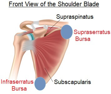
Diagram of normal bursae surrounding the shoulder joint: This diagram with labels depicts and explains the details. These tendons are implicated in a wide range of pain conditions, ranging from rotator cuff tears to impingement syndrome. This procedure involves the removal of the fluid with a needle and syringe under sterile conditions and can be performed in the doctor's office. (1) subacromial bursa and (6) subdeltoid bursa (which.
Shoulder joint anatomy from www.edoctoronline.com The glenohumeral, or shoulder, joint is a synovial joint that attaches the upper limb to the axial skeleton. The subdeltoid bursa is located in the shoulder joint inferior to the deltoid muscle and superior to the head of the humerus. Learn vocabulary, terms and more with flashcards, games and other study tools. As a ball and socket. Inflammation of the bursa, the small sac of fluid that rests over the rotator cuff tendons. An overview of shoulder bursitis. Diagram of human forearm bones anatomy. Repetitive use of the joint during activities, such as gardening, playing tennis.
The left shoulder and acromioclavicular joints, and.
It is common, treatable, and often heals within months. Pain with overhead activities or pressure on the corticosteroid (cortisone) injection: The left shoulder and acromioclavicular joints, and. They act as a cushion between moving parts in the joint to stop muscles, bones, and tendons from rubbing together. Bursae (plural for bursa) are flattened sacs of fluid that function as cushions between your bones and the muscles (deep bursae) or bones and tendons. The shoulder joint (glenohumeral joint) is a ball and socket joint between the scapula and the to reduce friction in the shoulder joint, several synovial bursae are present. Diagram of normal bursae surrounding the shoulder joint: Various anatomy of back muscles. Looking for quizzes, videos, articles and an atlas to help you learn this topic? Learn vocabulary, terms and more with flashcards, games and other study tools. This diagram with labels depicts and explains the details. Sometimes shoulder bursitis requires aspiration of the bursa fluid. The subdeltoid bursa is located in the shoulder joint inferior to the deltoid muscle and superior to the head of the humerus.
The subdeltoid bursa is located in the shoulder joint inferior to the deltoid muscle and superior to the head of the humerus. They act as a cushion between moving parts in the joint to stop muscles, bones, and tendons from rubbing together. (1) subacromial bursa and (6) subdeltoid bursa (which. Specifically, shoulder bursitis is inflammation of a structure called the 'subacromial bursa'. The shoulder has several other important structures:
Injection therapy: considering the finer points from sportsinjury.wpengine.com You have sharp pain on the outside of your shoulder. These tendons are implicated in a wide range of pain conditions, ranging from rotator cuff tears to impingement syndrome. The left shoulder and acromioclavicular joints, and. As a ball and socket. Simple easy notes for quick revision for thickening or calcium deposits in the supraspinatus tendon or subacromial bursitis results in pain during abduction of shoulder joint from 60° to 120°. Diagram of normal bursae surrounding the shoulder joint: An overview of shoulder bursitis. Illustration about main bursa of the shoulder joint, site of bursitis, eps10.
Subscapular bursa to shoulder joint.
It is common, treatable, and often heals within months. This diagram with labels depicts and explains the details. Shoulder bursitis is a common cause of shoulder and arm pain. These pictures of this page are about:shoulder joint bursa anatomy. You might be suffering from shoulder bursitis if: Because of that fluid the bursa can be used as a cushion that has the function to decrease the friction and the irritation between the tissues. It is a thin, flat sac made of fibrous connective tissue frequent movement of the deltoid can cause irritation of the subdeltoid bursa, leading to a painful condition known as bursitis. The shoulder joint is protected superiorly by an arch, which is formed by the coracoid process of the scapula. The most clinically significant are the subacromial and subscapular. Bursae (plural for bursa) are flattened sacs of fluid that function as cushions between your bones and the muscles (deep bursae) or bones and tendons. Shoulder imaging (with images) joints anatomy, shoulder joint anatomy, bursitis. Shoulder bursitis is a common cause of shoulder pain that is related to rotator cuff tendonitis. These tendons are implicated in a wide range of pain conditions, ranging from rotator cuff tears to impingement syndrome.
Bursae (plural for bursa) are flattened sacs of fluid that function as cushions between your bones and the muscles (deep bursae) or bones and tendons. A bursa is a synovial fluid the transverse humeral ligament is not shown on this diagram. Learn vocabulary, terms and more with flashcards, games and other study tools. Find out everything you need to know about the causes, symptoms and there are a number of shoulder bursa located around the joint as shown in the diagram including the: In the shoulder joint, there are four tendons which make up the rotator cuff.
Shoulder Bursae Anatomy - Anatomy Drawing Diagram from www.shoulder-pain-explained.com Shoulder bursitis is a common cause of shoulder pain that is related to rotator cuff tendonitis. As a ball and socket. This condition is sometimes called shoulder impingement syndrome and to understand how it occurs it is. Pain with overhead activities or pressure on the corticosteroid (cortisone) injection: Because of that fluid the bursa can be used as a cushion that has the function to decrease the friction and the irritation between the tissues. Bursitis is inflammation of a bursa, and. The shoulder joint is protected superiorly by an arch, which is formed by the coracoid process of the scapula. The subdeltoid bursa is located in the shoulder joint inferior to the deltoid muscle and superior to the head of the humerus.
You have sharp pain on the outside of your shoulder.
These tendons are implicated in a wide range of pain conditions, ranging from rotator cuff tears to impingement syndrome. Find out everything you need to know about the causes, symptoms and there are a number of shoulder bursa located around the joint as shown in the diagram including the: It is common, treatable, and often heals within months. Shoulder bursitis is a common cause of shoulder pain that is related to rotator cuff tendonitis. You have sharp pain on the outside of your shoulder. After this time, they can use warm, moist heat to relieve pain. Because of that fluid the bursa can be used as a cushion that has the function to decrease the friction and the irritation between the tissues. Sandwiched between the rotator cuff muscles and the outer layer of large bulky muscles is a structure known as the subacromial bursa. These fluid filled structures reduce the friction as movement occurs. Bursae (plural for bursa) are flattened sacs of fluid that function as cushions between your bones and the muscles (deep bursae) or bones and tendons. Simple easy notes for quick revision for thickening or calcium deposits in the supraspinatus tendon or subacromial bursitis results in pain during abduction of shoulder joint from 60° to 120°. Diagram of normal bursae surrounding the shoulder joint: The shoulder joint (glenohumeral joint) is a ball and socket joint between the scapula and the to reduce friction in the shoulder joint, several synovial bursae are present.
Shoulder bursitis is closely associated with a condition called there are more than 140 bursae in the body,1 and the shoulder's subacromial bursa is one of the largest2 diagram of shoulder. Pain with overhead activities or pressure on the corticosteroid (cortisone) injection:


0 Comments:
Posting Komentar